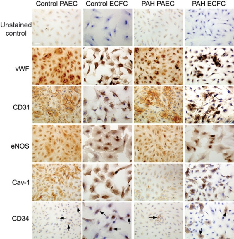
Figure 6.

ECFC express endothelial cell-specific markers. Representative images are shown. Both PAEC and ECFC demonstrate positive staining for CD31, vWF, cav-1, eNOS, and CD34. vWF stains intracellularly in both PAEC and ECFC, as expected for endothelial cells. CD31 exhibits staining localized to the membrane, particularly at sites of cell-cell contact, and in the cytosol. The cell-junctional staining for CD31 staining in ECFC is not as pronounced owing to cells not being confluent. eNOS staining is present in both PAEC and ECFC, particularly in the perinuclear region. Cav-1 stains both PAEC and ECFC. There is also CD34 staining in both PAEC and ECFC in both control and PAH groups (arrows). There are no significant differences in staining pattern between PAH and control groups. Scale bar is 50 μm.
