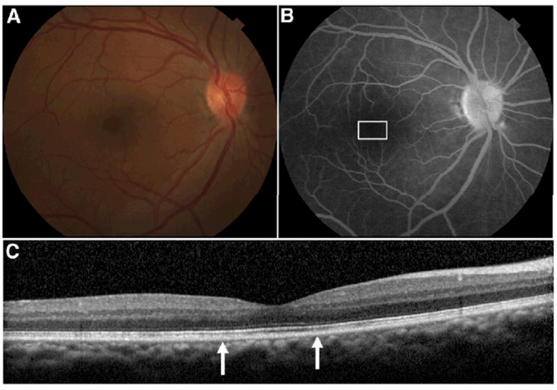
Figure 1. Clinical imaging, right eye.

A. Color fundus photo showing no macular abnormalities. B. Late frame fluorescein angiogram showed no window defect, staining or leakage. White box indicates area imaged by adaptive optics scanning ophthalmoscope (AOSO) as seen in figure 2. C. Spectralis SD-OCT horizontal scan through the fovea showing no outer retinal abnormalities. Area between arrows indicates region imaged by AOSO as seen in figure 2.
