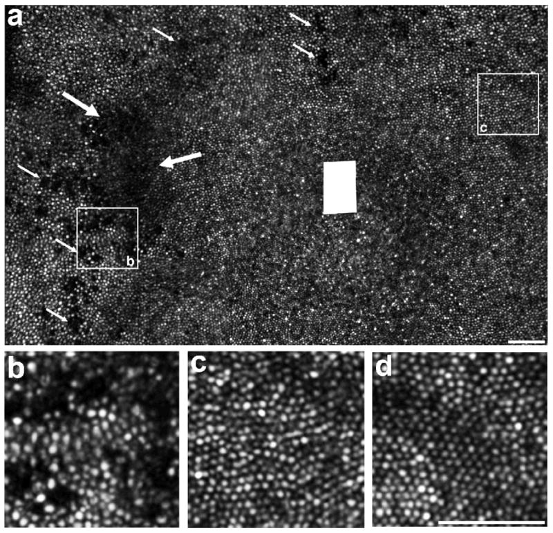
Figure 2. Disrupted photoreceptor mosaic, macula, right eye.

A. Adaptive optics scanning ophthalmoscope montage shows a large, crescent-shaped area of photoreceptor disruption (edges indicated by large arrows) temporal to fovea. Multiple other areas of photoreceptor disruption are also present (small arrows). Foveal center was not imaged, and is marked by the solid white rectangle. B. Magnified view of a patch of retina 1-degree temporal from the fovea, centered on an area of significant photoreceptor disruption. C. Magnified view of a patch of retina 1-degree nasal from the fovea, showing regularly packed cone photoreceptor mosaic. D. Image from a normal control, about 1-degree temporal from the fovea. Scale bar for all images is 50 microns.
