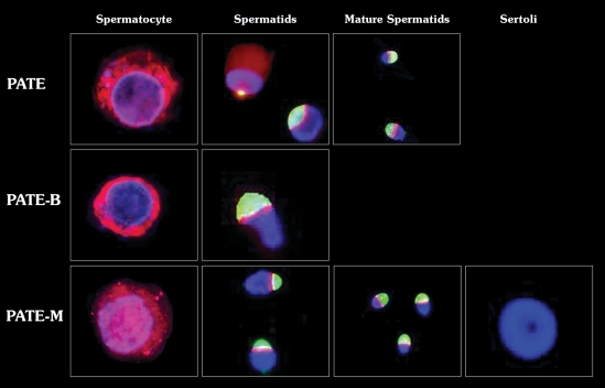
Figure 2.
PATE, PATE-B and PATE-M proteins localization in germ cells isolated from human testicular tissue. PATE proteins were stained with rhodamine-conjugated secondary Ab (red), the acrosome with Pisum sativum agglutinin fluorescein isothiocyanate (PSA–FITC, green) and the nucleus with 4′,6-diamidino-2-phenylindole DAPI (blue). The percentage of stain cells was 85, 50 and 2% for PATE, PATE-M and PATE-B proteins, respectively. Staining of PATE and PATE M proteins was detected in the cytoplasm of spermatocytes and spermatids and around the acrosome in different stages of spermatids. PATE-B protein was observed in the cytoplasm of spermatocytes and around the acrosome of the spermatids. Magnification ×1000.

