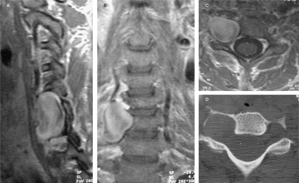
Figure 5.
Case 4. On T2W MRI sequences enlargement of transverse foramen due to a mass presumably representing a large aneurysm (high signal due to low flow and turbulence) can be seen that demonstrates contrast enhancement (A-C: T1 weighted images after contrast) and bone erosion on CT (D). Adjacent structures are compressed.

