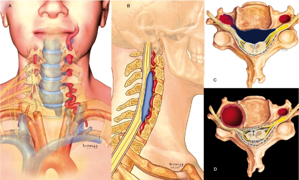
Figure 7.
Schematic Illustration of the pathophysiology of the presented cases. In the first three patients, a vertebrovertebral arteriovenous fistula was present. In these drawings, the dysplastic artery ruptured to a venous vertebral compartment and major important reflux to the epidural venous plexus causing spinal cord and radicular compression can be appreciated (AC). D) Illustration of the vertebral aneurysm with irregular and thick walls of the RVA associated with adjacent bone erosion from the pulsatility and enlargement of the transverse foramen causing compression of the exiting nerve root as was present in patient 4.

