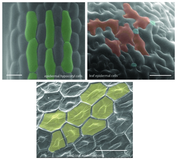
Figure 1.
Scanning electron microscopy (SEM) micrographs showing variation in epidermal cell size and shape in different plant organs. Etiolated hypocotyls, leaves and seeds from Arabidopsis were sputter coated with gold-palladium using Hummer VI sputtering system (Anatech) and visualized using Hitachi-S-800 SEM. Scale bars = 30 μm.

