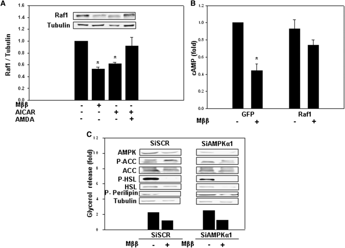
Fig. 5.
Suppression of Raf1 expression by Mββ and AICAR. 3T3-L1 adipocytes or COS-1 cells were incubated for 24 h in the presence of 350 μM or 250 μM Mββ, respectively, 750 μM AICAR and 25 μM AMDA as indicated, and in the absence or presence of 100 nM isoproterenol (Iso), 30 μM IBMX or 10 μM forskolin added to the incubation medium 1 h prior to sampling as indicated. Lipolysis (C) was quantified by glycerol release as described in Experimental Procedures. P-HSL, HSL, P-perilipin, AMPK, P-ACC(S79), ACC, Raf1, and tubulin (A, C) were determined by SDS-PAGE/Western blot analysis as described in Experimental Procedures. Cellular cAMP content (B) was determined as described in Experimental Procedures. A: Suppression of Raf1 by Mββ and AICAR in 3T3-L1 adipocytes. Raf1 protein of nontreated cells is defined as 1.0. Mean ± SE of three independent experiments. *Significant compared with nontreated cells (P < 0.05). Inset: Representative blot. B: Rescue of Mββ-suppressed cAMP levels by overexpressed Raf1 in COS-1 cells. COS-1 cells were transfected with GFP-Raf1 expression vector or with the empty GFP plasmid as indicated, and were further cultured in the presence of forskolin/IBMX. cAMP values were corrected for transfection efficiency, estimated by the percentage of GFP expressing cells. Cellular cAMP content of nontreated controls transfected with the empty GFP plasmid is defined as 1.0. Mean ± SE of four independent experiments. *Significant compared with nontreated GFP-transfected cells (P < 0.05). C: Mββ suppression of isoproterenol-induced lipolysis, P-HSL, and P-perilipin prevails in AMPK-deficient 3T3-L1 adipocytes. 3T3-L1 adipocytes were transfected with siAMPK or scrambled Si (SCR). Representative Western blots and lipolysis (filled bars) of cells transfected with siAMPK or scrambled Si. Glycerol release of nontreated cells is defined as 1.0. Representative experiment.

