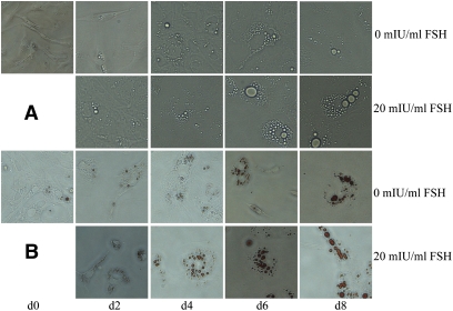
Fig. 5.
Morphological changes and lipid deposition induced by 20 mIU/ml FSH in preadipocytes during differentiation in vitro (inverted microscope, 200×). A: The change from cells being spindle-shaped to larger oval or rounded forms was accelerated in those exposed to 20 mIU/ml FSH when compared with those not treated with FSH. B: Lipid, stained with Oil red O, accumulated as fewer but much larger locules in cells exposed to 20 mIU/ml FSH when compared with those not treated with FSH.

