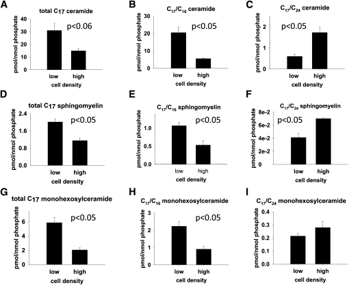
Fig. 4.
Metabolic labeling with C17 sphingosine of neuroblastoma cells cultured at low and high cell densities. SMS-KCNR cells were cultured at low (50%) and high (90%) cell densities. After 48 h, cells were labeled with 1 µM C17 sphingosine for 30 min. Lipids were extracted, and C17 sphingolipid species were measured by LC/MS analyses. C17 sphingolipid levels were normalized to cellular lipid phosphate. Error bars represent the results from three independent experiments. (A) Total C17 ceramide (the data used for calculating the total C17 ceramide is shown in supplementary Table I). (B) C17/C16 ceramide. (C) C17/C24 ceramide. (D) Total C17 sphingomyelin (the data used for calculating the total C17 sphingomyelin is shown in supplementary Table II). (E) C17/C16 sphingomyelin. (F) C17/C24 sphingomyelin. (G) Total C17 monohexosylceramide (the data used for calculating the total C17 monohexosylceramide is shown in supplementary Table III). (H) C17/C16 monohexosylceramide. (I) C17/C24 monohexosylceramide.

