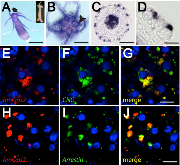
Figure 2.
Studies of gene expression suggest a role for opsin-mediated photosensitivity in the regulation of cnidocyte discharge. (A-D) Colorimetric in situ hybridization with HmOps2 probe. Opsin expression is strongly localized to the hypostome (arrow), tentacles, and the ring-like ganglion (arrow head) that surrounds the mouth (A and B). (C and D) Cryosection of tentacle reveals HmOps2 expression in non-cnidocyte cell types. Cnidocytes capsules are clearly visible in these preparations. (E-I) Confocal fluorescence in situ hybridization shows that opsin transcripts co-localize with CNG (E-G) and arrestin (H, I) in battery complexes of the hydra. Blue cells with dark centers are the capsules of stenotele cnidocytes, which stain with DAPI [62]. Signal in (E-I) and (E-G) is located at different focal planes relative to central stenotele cnidocytes. Inset of (A), sense riboprobe negative control. Scale bars in (A) = 1 mm, in (B) = 500 μm, in (C) = 100 μm, in (D) and (E-J) approximately 30 μm.

