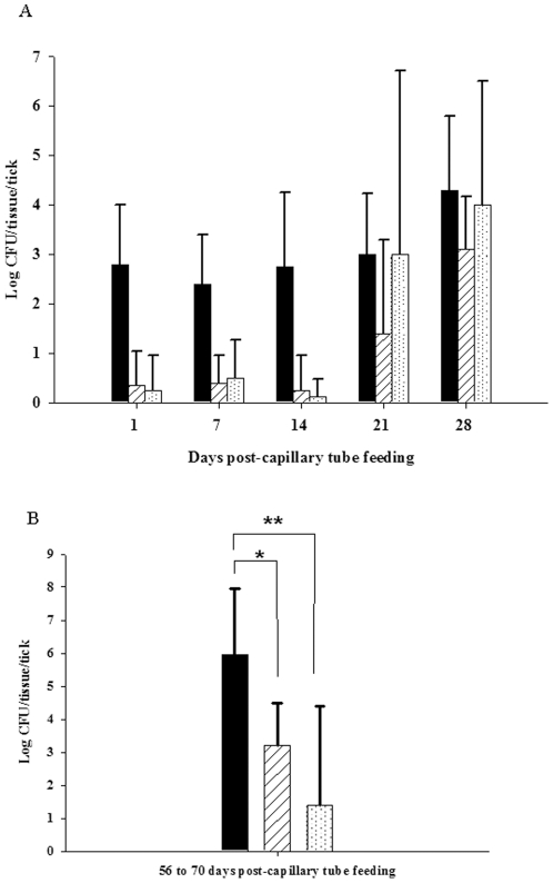
Figure 5. Tissue dissemination of LVS in adult D. variabilis.
(A) Solid black bar- gut, white bar with diagonal lines- salivary gland, white bar with dots- hemolymph. For each time point the n was 5. Error bars indicate standard deviation. (B) Tissue dissemination of LVS in adult D. variabilis between 56 and 70 days post-capillary tube feeding. Solid black bar - gut, white bar with diagonal lines - salivary glands, white bar with dots - hemolymph. For each time point, n was 10. Error bars indicate standard deviation. * Unadjusted P = 0.008, ** unadjusted P<0.001.

