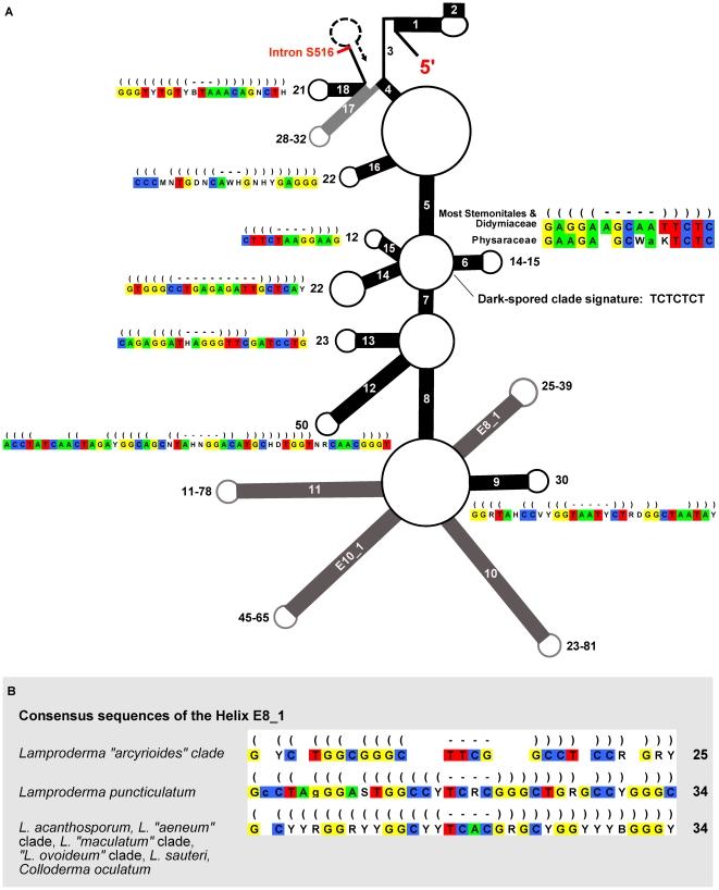
Figure 4. Secondary structure and signatures.
A: Schematic secondary structure of the first part of the SSU rRNA (after [51]), encompassing the first 18 helices and part of the 19th, up to the first intron insertion site. Helices (paired strands) common to all domains of life are represented by black rectangles and numbered (E8_1, E10_1 are unique to eukaryotes), lengths are given next to the loops. Single-stranded segments are indicated by thin lines. For helices whose length is conserved, a 95% consensus is shown. Helices whose length is variable are in grey and the length range is given. Consensus sequences of the helices are shown in the Stockholm format (parentheses = stem, hyphens = loop, blank = no paring); lower-case characters indicate a base that is not present in all sequences of the group. B: Consensus sequences of the helix E8_1 of several Lamproderma clades, showing high variability and clade-specific sequence signatures. Lengths are more conservative than sequences.

