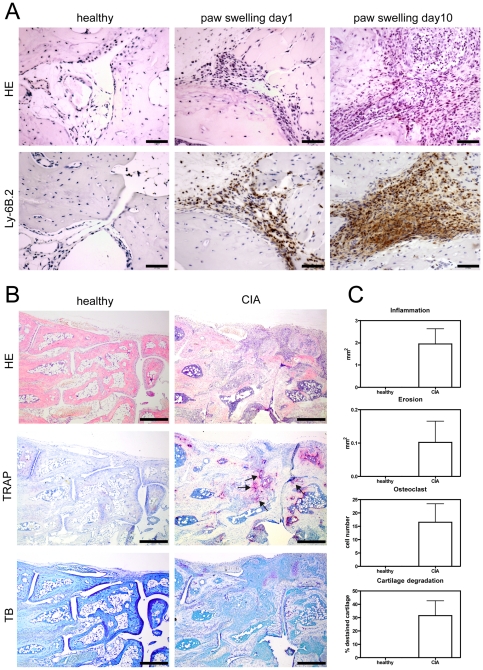
Figure 1. Analysis of GMC migration and histomorphological assessment of arthritis in animals with CIA.
A) Paraffin sections were stained with HE and anti-Ly-6B.2 mAb. In contrast to healthy animals GMC cells can be detected in animals with CIA at the earliest time-points (paw swelling day 1) of clinical signs of arthritis and are abundant at later (paw swelling day 10) time-points. Scale bars represent 50 µm. Representative pictures are shown from experiments that were performed in three healthy animals and three animals with CIA. B) Representative samples of paraffin sections from hind paws of three healthy control animals and three animals with CIA stained for HE, TRAP and TB are shown. Arrows point to areas of bone erosions. Scale bars represent 500 µm. C) Histomorphological signs of arthritis were quantified in three CIA subjects (day 11±6 of paw swelling) as compared to three healthy control subjects. Areas of inflammation, bone erosions, osteoclasts and cartilage degradation can be detected in CIA subjects but not in healthy control animals. Graph bars represent mean values ± s.e.m.

