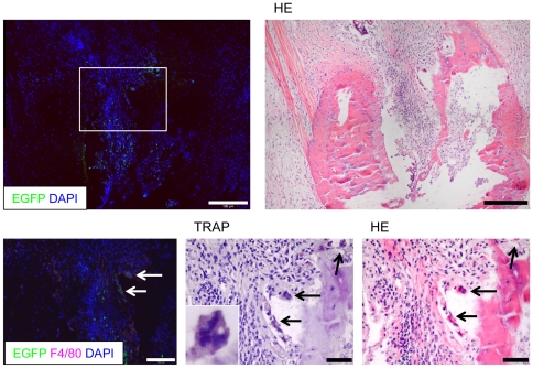
Figure 5. Combined sequential immunofluorescence/histomorphological analysis of monocytes/osteoclasts.
Analysis of cryo sections stained with DAPI (left) and subsequent HE (right) staining (upper two pictures). A higher magnification of the insert demonstrates that individual EGFPlow cells (arrows) express F4/80 and TRAP, are multinucleated and localized in close vicinity to the bone tissue. A higher magnification insert in the middle pictures shows one EGFPlowF4/80+TRAP+ cell. Representative pictures are shown from experiments that were performed in three animals with CIA. Scale bars represent 100 µm.

