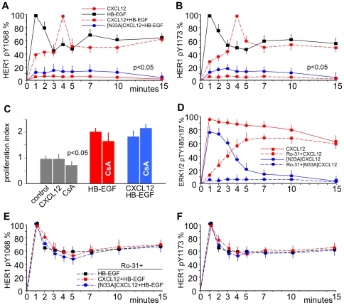
Figure 3. CXCL12 regulates HER1 Y1068 and Y1173 phosphorylation via G-proteins.
5637 or HeLa cells were stimulated with 25 ng/mL HB-EGF or with 200 ng/mL CXCL12 or N33A]CXCL12 alone or followed after 1 minute by 25 ng/mL HB-EGF. HER1 Y1068 or ERK1/2 TY185/187 phosphorylation was evaluated at the indicated time-points by using specific mAbs and was expressed as percentages of phosphorylation at the specified times after normalization as phosphorylated molecule/total molecule ratios. (A) HER1 phosphorylation at Y1068 in HeLa cells induced by HB-EGF alone (black) was abolished by prestimulation with [N33A]CXCL12 (blue) and modified by prestimulation with CXCL12 (red): maximum phosphorylation was reached at 4 minutes, and the plateau after the initial spike was around 50% of the maximum at 10 minutes. No phosphorylation was induced by CXCL12 alone. (B) Phosphorylation at Y1173 in 5637 cells displayed the same kind of pattern. (C) Prestimulation with CXCL12 did not modify the mitogenic effect of HB-EGF. (D) Stimulation with CXCL12 induced ERK1/2 phosphorylation (red) resulting from two spikes: a G-protein-dependent (blue) and a β-arrestin-dependent (red) phosphorylation spike. By using the PKC inhibitor Ro-31 the G-protein-dependent spike was abolished, whereas the β-arrestin-dependent spike persisted. [N33A]CXCL12 induced only the G-protein-dependent spike (blue), which was abolished by Ro-31. (E) Ro-31 abolished the effects of prestimulation with CXCL12 or [N33A]CXCL12 at Y1068 in 5637 cells. (F) The same pattern at Y1173 in HeLa cells. The means ±SD of 10 experiments are depicted.

