Figure 2.
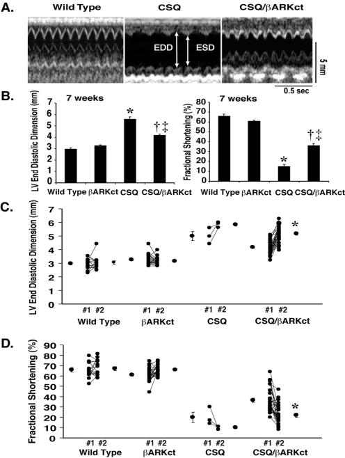
Analysis of cardiac function by noninvasive echocardiography in conscious mice. (A) Transthoracic M-mode echocardiographic tracings in 7-week-old wild-type (Left), CSQ (Center), and CSQ/βARKct (Right) mice. LV dimensions are indicated with the double-sided arrows. EDD, end diastolic dimension; ESD, end systolic dimension. Wild-type mice have normal chamber size, whereas the CSQ mice have chamber dilation and depressed cardiac function. The CSQ/βARKct mice have only moderate chamber dilation and slightly reduced cardiac function as compared with the wild-type mice. (B) Echocardiographic findings in 7-week-old wild-type and transgenic mice. LV EDD (Left) and percent fractional shortening (Right) are shown. Wild type, n = 15; βARKct, n = 23; CSQ, n = 14; CSQ/βARKct, n = 31. *, P < 0.0001, CSQ vs. wild type; †, P < 0.0001, CSQ/βARKct vs. CSQ; ‡, P < 0.0001, CSQ/βARKct vs. wild type. (C) Data from serial echocardiograms in the same mouse at 7 weeks (#1) and 11 weeks (#2) of age for LV EDD. (D) Percent fractional shortening in the same mouse. Wild type, n = 14; βARKct, n = 22; CSQ, n = 3; CSQ/βARKct, n = 28. *, P < 0.001, CSQ/βARKct (#2) vs. CSQ/βARKct (#1).

