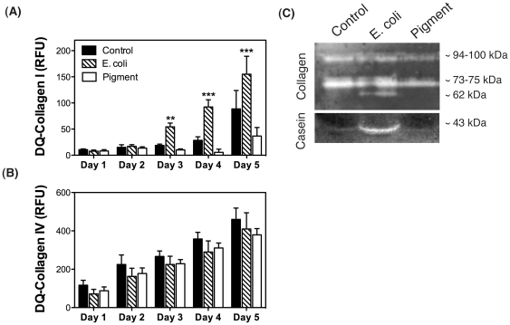
Figure 7. Collagenolytic activity of porcine TM cells phagocytically challenged to E.coli or pigment in the presence of the self-quenched fluorescent substrates DQ-Collagen I (A) or DQ-Collagen IV (B).
Values represent mean ± SD. **p<0.005, ***p<0.0005 (t-test, n = 3). (C) Collagenolytic and caseinolytic activities in the culture media of porcine TM cells phagocytically challenged to E.coli or pigment evaluated by in-gel collagen I or casein zymography. Lytic activity is shown as clear bands. Zymograms are representative from three independent experiments.

