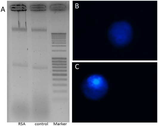
Figure 4. Effect of RSA to induce apoptosis in Drosophila melanogaster S2 cells.
(A) DNA fragmentation in S2 cells. Cells were treated with 0.7 µM RSA compared to control (untreated) cells. Ten micrograms of extracted DNA was loaded on the 2% agarose gel. (B,C) Nuclear condensation assay: Upon treatment with 0.7 µM RSA, the nuclei of the S2 cells were stained with Hoechst. Typically, treated cells (C) showed a normal, non-fragmented nucleus similar to the untreated control cells (B).

