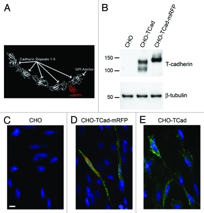
Figure 3. Construction of tagged T-cadherin. (A) Prediction of T-cadherin structure, showing insertion of schematic 3xGly-mRFP-3xGly peptide folding separately in between cadherin motifs four and five. (B) Western blot of CHO cells with or without transfection with wild type T-cadherin or the Tcad-mRFP construct. (C–E) T-cadherin (green) immunostaining performed on living non-permeabilized cells, illustrating Tcad surface localization. Tcad-mRFP fluorescence-red. Scale bar: 10 μm. (C) Wild type CHO cells. (D) CHO cells transfected with Tcad-mRFP. (E) CHO cells transfected with wild type T-cadherin. Note that some Tcad-mRFP is detected in the cytoplasm (D), which probably reflects protein during processing and transport.

An official website of the United States government
Here's how you know
Official websites use .gov
A
.gov website belongs to an official
government organization in the United States.
Secure .gov websites use HTTPS
A lock (
) or https:// means you've safely
connected to the .gov website. Share sensitive
information only on official, secure websites.
