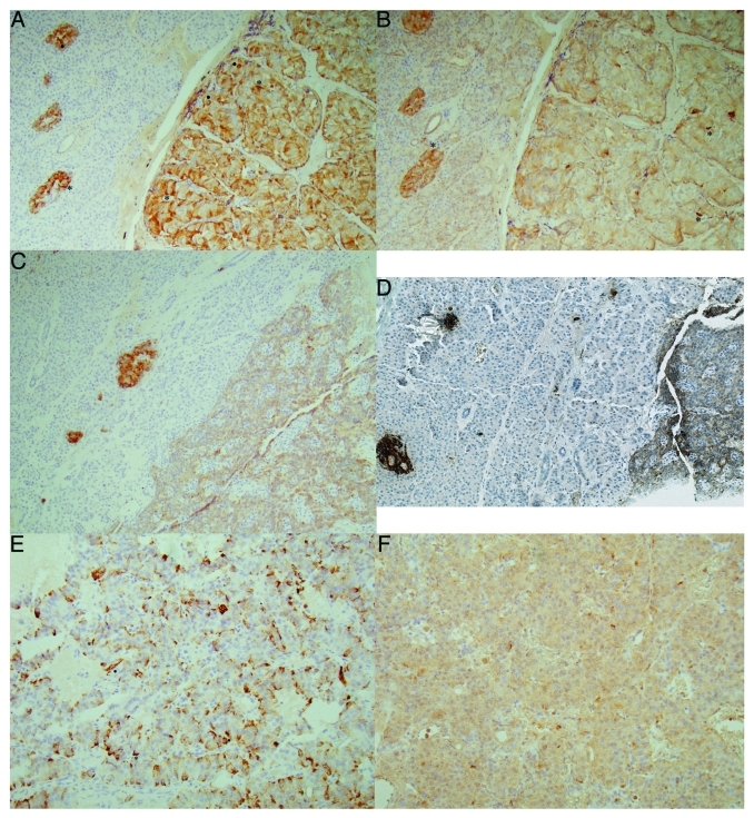
Figure 2. Insulinoma cases. Case 1 (A and B), Case 2 (C and D) and Case 3 (E and F). Case 1, benign insulinoma, revealed diffusely strongly positive staining for insulin (A) and diffusely moderately positive staining for IAPP (B) with scattered dense staining for insulin (.), some of which were dense sickle-shaped cytoplasm, often showing no nucleus (*). Case 2, another benign insulinoma, was moderately positive for insulin (C) and weakly positive for IAPP. A double immunostianing for insulin (brown) and IAPP (blue) revealed the same tumor cells positive for both insulin and IAPP (D). Case 3, malignant insulinoma, revealed diffusely moderately positive staining for insulin (E) and diffusely weak staining for IAPP (F) with some scattered strongly positive cells for insulin (.) only. (A, C and E) Insulin, (B and F) IAPP, (D) a double immunostained for insulin (brown) and IAPP (blue).

An official website of the United States government
Here's how you know
Official websites use .gov
A
.gov website belongs to an official
government organization in the United States.
Secure .gov websites use HTTPS
A lock (
) or https:// means you've safely
connected to the .gov website. Share sensitive
information only on official, secure websites.
