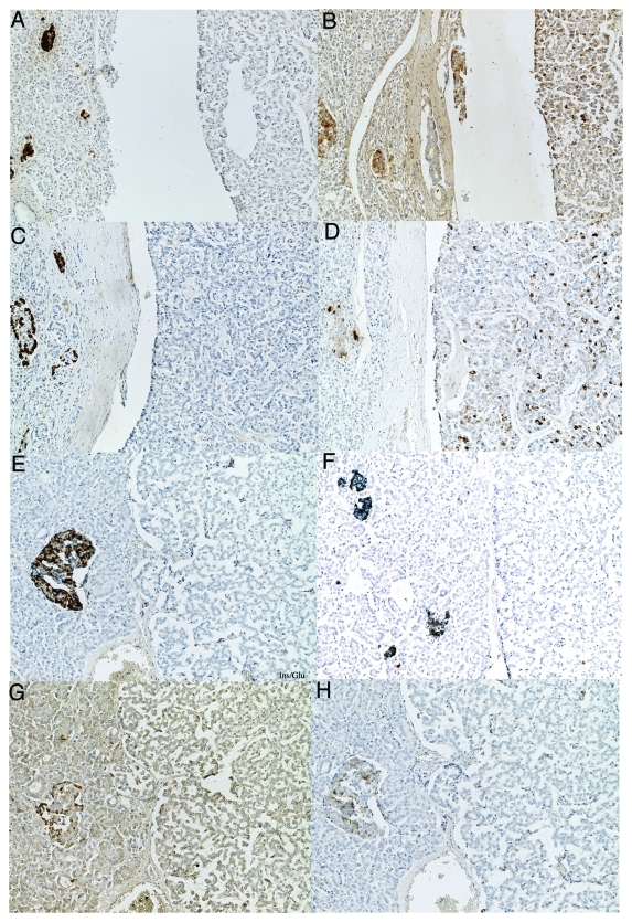
Figure 3. IAPP-positive PPoma (A–D) and IAPP-negative non-functioning tumor (E–H). This PPoma revealed many strongly PP positive cells (D) and scattered moderately IAPP positive cells (B) whereas tumor cells were negative for insulin (A), glucagon (C) and SRIF. This non-functioning tumor was negative for IAPP (F and H) and was also negative for insulin (E and F), glucagon (E), SRIF (G) and PP (H). (A) Insulin, (B) IAPP, (C) glucagon, (D) PP, (E) insulin/glucagon double stained, (F) IAPP/Insulin double stained, (G) SRIF, (H) IAPP/PP double immunostained.

An official website of the United States government
Here's how you know
Official websites use .gov
A
.gov website belongs to an official
government organization in the United States.
Secure .gov websites use HTTPS
A lock (
) or https:// means you've safely
connected to the .gov website. Share sensitive
information only on official, secure websites.
