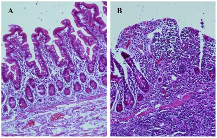
Figure 7. Histological characteristics of the ileal mucosa from CD patient.
H&E staining showed normal epithelial architecture and few infiltrated inflammatory cells in the nonulcerated tissues (A). The mucosal specimen from ulcerated ileum revealed the disrupted mucosal architecture and the infiltration of inflammatory cells in (B). Magnifications: ×100.

