
Figure 2. Constitutive bodies and dynamic bodies.
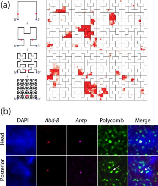
(a) Hilbert folding representation of Polycomb binding sites on chromosome 3R of Drosophila. Some 75-100 (depending on how they are counted) PC binding sites correspond to this map that shows a high degree of clustering along the chromosome. The diagrams on the left explain how the iterative Hilbert folding (for two dimensions, called Peano folding; for applications to genomic data see ref. 51) of the linear chromosome arm is achieved. Figure kindly provided by P. Kharchenko. (b) The Drosophila Antp gene (magenta) and the Abd-B gene (red) visualized by FISH co-localize within a PcG body (Polycomb) in nuclei from the embryonic head region, where both are repressed by PcG complexes. In nuclei from the posterior region, the active Antp gene moves out of the PcG body but the repressed Abd-B gene remains associated with the PcG body (reproduced from ref. 10**, with permission)