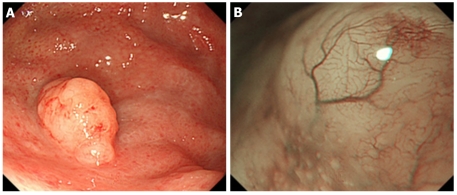
Figure 5.
Endoscopic findings of gastric mucosa-associated lymphoid tissue lymphoma. A: An elevated lesion, 10 mm in diameter, was observed in the greater curvature of the gastric fornix; B: Narrow band imaging findings. The mucosa that had lost its glandular structure, appeared shiny and had a tree-like appearance.

