Figure 5.
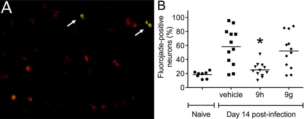
Effects of the indole enantiomers, 9g and 9h, on neuronal survival in the brains of NSV-infected mice. The anatomically-defined hippocampus, a site of heavy NSV infection, was chosen for further study. (A) Representative fluorojade staining of damaged hippocampal neurons in the brains of NSV-infected mice. NisslRed was first used to label neurons in sections through the hippocampus, while fluorojade staining (arrows) was then performed to identify those cells undergoing active degeneration. (B) The proportion of fluorojade-positive hippocampal neurons was determined in quadruplicate slides prepared from 3 mice in each group. Statistical differences (*p<0.05) in the number of labeled (injured) neurons were determined by an unpaired Student’s t-test compared to vehicle-treated control animals.

