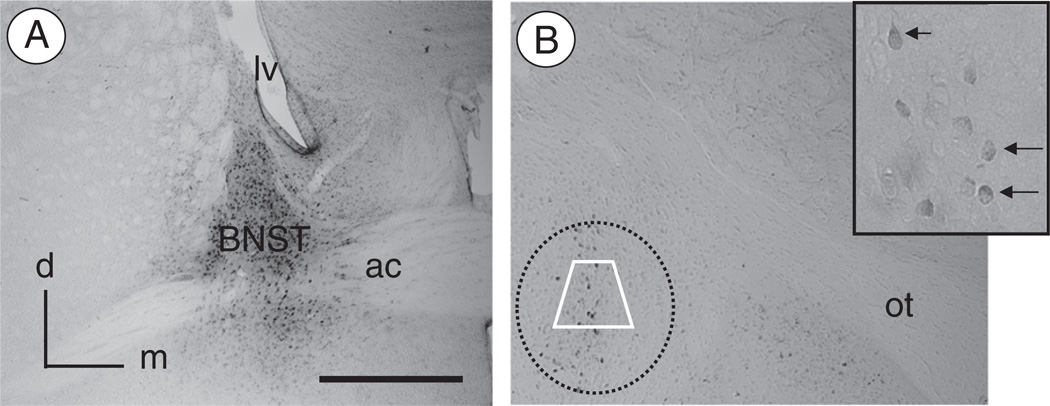
Fig. 1.
Injection of FG in the BNST results in retrograde labeling in CeA neurons(A). Light micrograph illustrating a representative example of FG immunolabeling in the BNST after local administration of the tracer. Immunoperoxidase reaction product for FG is present in the dorsal as well as ventral BNST surrounding the anterior commissure. There is little FG labeling in the regions adjacent to the BNST. (B). Retrogradely labeled neurons are present in the CeA. At high magnification (inset), immunoperoxidase reaction product for FG can be seen throughout the cytoplasm of neuronal cell bodies (arrows). Electron microscopic analysis of FG, NR1, and/or μOR labeling was performed in samples taken from the region of the CeA represented by the area bound by the trapezoid. ac: anterior commissure; d: dorsal; lv: lateral ventricle; m: medial; ot: optic tract. Scale Bar: 1 mm.

