
Figure 1. Neuroimaging methods relevant to the assessment of cognitive changes.
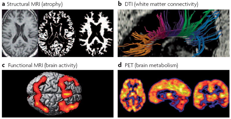
a ∣ Structural magnetic resonance imaging (MRI) provides a high resolution picture of normal neuroanatomic details and atrophy (T1-weighted scans), and visible pathology such as microvascular and inflammatory lesions (T2-weighted scans; fluid-attenuated inversion recovery (FLAIR) scans). Semi-automated methods can be used to segment or classify the structural images into the main tissue compartments, including grey and white matter, cerebrospinal fluid and hyperintense lesions, which reflect microvascular changes or areas of demyelination. Software is available to quantify the volume and other characteristics of each tissue type. b ∣ Diffusion tensor imaging (DTI) is a recently developed technique that can be used to assess pathological changes in grey matter (increased mean diffusivity) and the loss of integrity of white matter fibre bundles (decreased fractional anisotropy). Tractography software enables the identification of directional fibre bundles such as subregions of the corpus callosum (shown). c ∣ Functional MRI (fMRI) uses blood-oxygen-level dependent (BOLD) contrast or perfusion measurements to assess the functional activation of cortical and subcortical regions during the performance of cognitive or sensorimotor tasks in the scanner. Bilateral frontal and parietal activation can be seen, representing the mean activation observed in a group of healthy individuals performing a working memory task. d ∣ Positron emission tomography (PET) using the fluorodeoxyglucose (FDG) radiotracer provides a measure of neuronal metabolism. Other applications of molecular imaging methods such as PET provide data on cerebral blood flow or specific neurotransmitter–receptor systems. Most of these methods have not yet been examined in systematic, prospective studies of chemotherapy-induced or cancer-associated cognitive changes, but these approaches hold promise for identifying the neural bases of such changes.