
Figure 3.
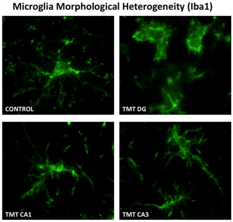
Heterogeneity of microglia responses within various hippocampal regions were detected at 72 hrs post-TMT (2mg/kg) injection. In the hippocampus of normal mice, Iba-1 staining of microglia showed small cells with thin, lightly stained, ramified processes. Within the dentate granule cell layer, microglia displayed a more rounded morphology with retraction of processes consistent with an amoeboid phagocytic phenotype. Within the CA1 and the CA3 pyramidal cell layers, microglia showed hypertrophy of processes with indication of retraction. Microglia were detected by rabbit polyclonal antibody to ionized calcium-binding adaptor molecule 1 (Iba1; 1:500; 1h, RT; Wako Chemicals, Richmond, VA) detected with IgG Alexafluor 488 (1:1000, Molecular Probes). Digital images were acquired using a SpotRT™ cooled, charged-couple device camera (Diagnostic Instruments, Sterling Heights, MI) on a Leica DMRBE microscope (Wetzlar, Germany) equipped with epifluorescence and Z-control and Metamorph™ (Universal Imaging Co., Downingtown, PA). 100x image stacks were acquired, deconvolved and 3D reconstruction by maximum projection shown at 20 degree angle.