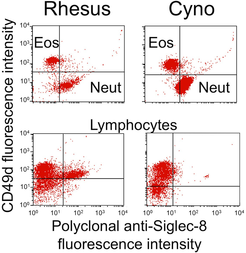
Fig. 3.
Polyclonal Siglec-8 labeling of eosinophils (Eos), neutrophils (Neut), monocytes (Mono), and lymphocytes (Lymph) in rhesus and cynomolgus monkey (Cyno) blood samples. Shown are results from a single experiment representative of three experiments. Note that the gating strategies for these experiments included light scatter to separate granulocytes from monocytes and lymphocytes, and then CD49d antibody labeling was used to distinguish eosinophils (positive) from neutrophils (negative)

