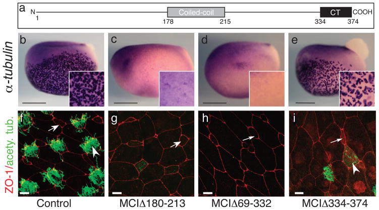
Figure 6. Domains required for MCI function.
(a) Diagram of MCI. (b–e) Shown are embryos injected with RNA encoding different MCI mutants as indicated, fixed at stage 13/14, and stained for α-tubulin RNA expression (f–i) Confocal images of the skin of embryos injected with RNA encoding different MCI mutants, fixed at stage 28, and stained with ZO-1 (red) and acetylated tubulin (green) antibodies to label cell boundaries and cilia, respectively. Scale bars=0.5mm (b–e), 10 microns (f–h), or 20 microns (i).

