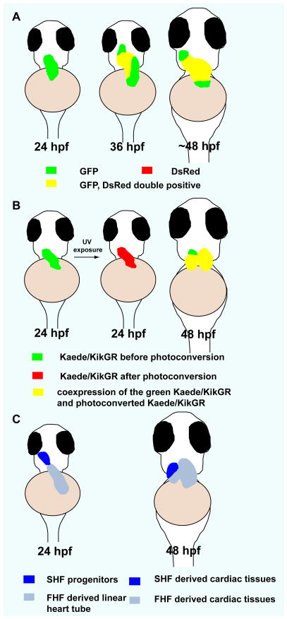
Fig 2.
Second heart field in zebrafish. A) Temporal order of cardiomyocyte differentiation: newly differentiated cardiomyocytes (green), earlier differentiated cardiomyocytes (yellow). B) Kaede/kikGR photoconversion experiments suggest that cardiomyocytes at the arterial pole of the zebrafish heart differentiate after the formation of the linear heart tube (green). C) Contribution of the SHF to zebrafish heart development. At 24 hpf, SHF progenitors are located immediately adjacent to the ventricle. At 48 hpf, SHF derived cells populate the distal segment of the ventricle as well as the outflow tract. Ventral views, anterior to the top. (adapted from de Pater E et al., Development. 136:1633–1641, Lazic and Scott, Developmental biology. 354:123–133, and Zhou Y et al., Nature. 474:645–648)

