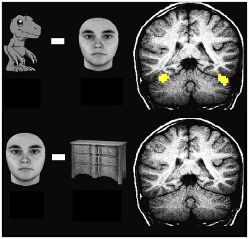
Figure I.

Activation of the FFA to Digimon (top panel), but not to faces (bottom panel) in patient DD. Right and left are reversed by radiological convention. Voxels are colored if the smoothed data have at ≥4 (which corresponds to P < .0001 uncorrected). Adapted from [145].
