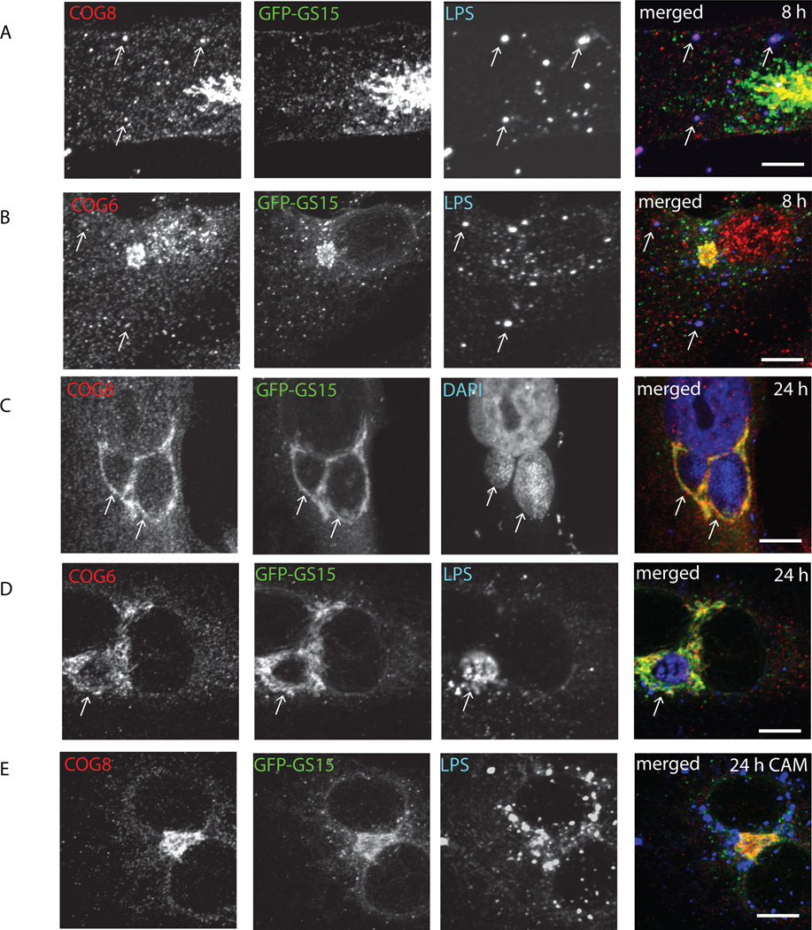
Fig. 5. COG complex subunits localized to chlamydial inclusions before GS15.
Stably transfected GFP-GS15 HeLa cells infected with C. trachomatis serovar L2 (A–C) or serovar D (D–E) were fixed either 8 h (A, B) or 24 h (C, D, E) after infection. In (E) cells were treated with chloramphenicol 3 h after infection to block bacterial protein synthesis. Cells were stained for chlamydial LPS (HiLyte Fluor-555) and COG8 (HiLyte Fluor-647) (A, E), DAPI and COG8 (HiLyte Fluor-647) (C), or chlamydial LPS (HiLyte Fluor-555) and COG6 (HiLyte Fluor-647) (B,D). The merge images are shown on the right. Arrows indicate the membrane of chlamydial inclusions. Scale bar, 10 µm.

