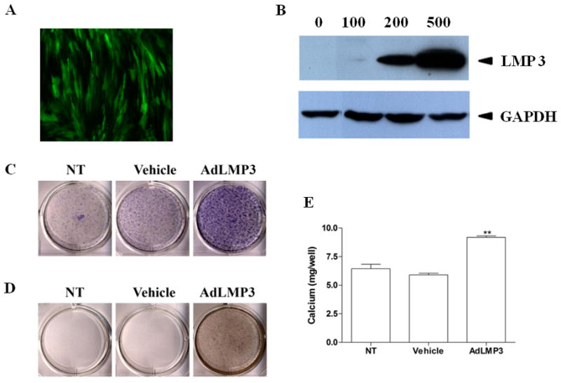
Figure 1. LMP3 promotes mineralization in PDL cells in vitro.

(A) PDL cells were transduced overnight with AdeGFP at MOI=200. The efficiency of transgene expression was evaluated by fluorescent invertoscope at 72h post-transduction. (B) PDL cells were transduced with AdLMP3 at different MOIs. Total protein lysate was harvested at day 3 and the exogenous LMP3 protein expression was analyzed by Western blot. LMP3 is approximately 16 kDa. (C) PDL cells were transduced with AdLMP3 or vector-only adenovirus. Cells were then induced to undergo osteogenic differentiation. At 1w, cells were fixed and ALP staining was performed. (D) At 2w, von Kossa staining was used to assess the matrix mineralization. (E) Extracellular calcium was measured (C). (*: p<0.05 compared to vehicle and NT; **: p<0.01 compared to Vehicle and NT; NT: no treatment)
