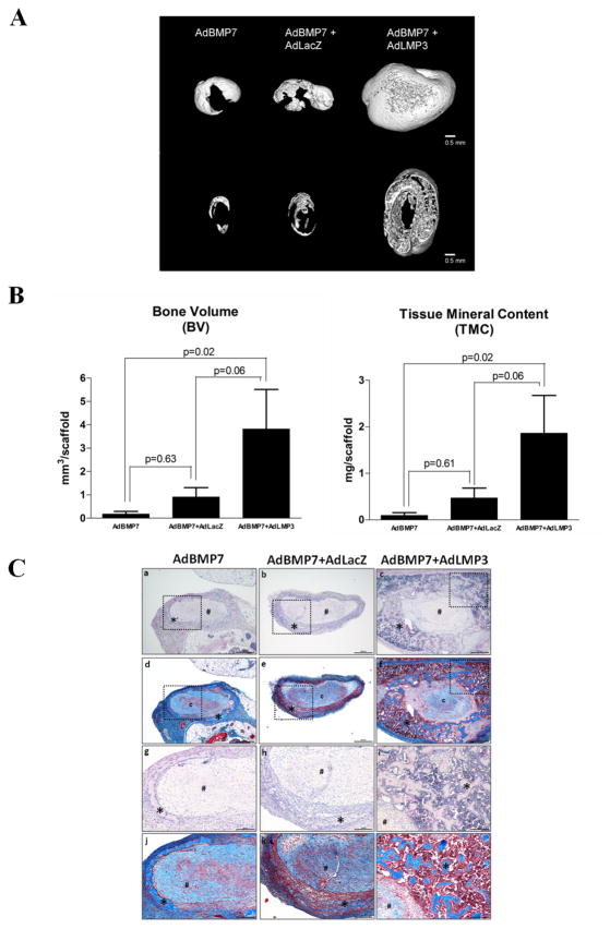
Figure 4. AdLMP3 gene transduction promotes bone formation synergistically with AdBMP7.
PDL cells were transduced with AdBMP7 (MOI 50), AdBMP7 (MOI 50)/LacZ virus or AdBMP7 (MOI 50)/AdLMP3 (MOI 200). After 24 h, 1×10⁶ cells were suspended into type I collagen scaffolds and subcutaneously implanted into immunodeficient mice. Implants were harvested and analyzed after 3 weeks. Samples were scanned by μ-CT. (A) Representative images of each group. Upper: 3D reconstruction. Lower: section view of the middle 1/3. (B) Quantitative analysis was performed to measure bone volume (BV) and tissue mineral content (TMC). (C) H&E (a–c, g–i) and Masson’s trichrome (d–f, j–l) stains were used to demonstrate the ectopic bone formation. a–f, low magnification, 40x. g–l: high magnification of the selected field (dot squared area), 100x. * represents bone and # represents collagen scaffold. n=8 in every group.

