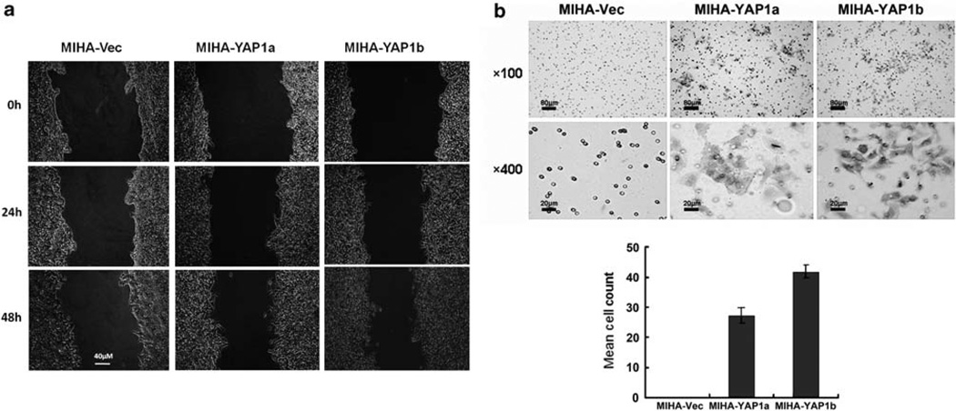
Figure 2.
YAP1 enhanced migration and invasion abilities of MIHA cells. (a) Wound healing assay. At 48 h, both MIHA-YAP1a and MIHA-YAP1b clones exhibited faster closure of the gap than MIHA-Vec cells did. Images were taken immediately after scratching the cultures 0 h, 24 h and 48 h later. Magnification: × 200, scale bar: 40 µm. (b) Matrigel invasion assay. Representative fields of invaded cells on the membrane are shown. Penetrated cells were stained with 0.1% crystal violet. MIHA-YAP1 clones were able to penetrate the Matrigel membrane after 72-h incubation. Histograms in the lower panel showed the mean cell counts of the invaded cells. Magnification × 100, scale bar: 80 µm; magnification: × 400, scale bar: 20 µm.

