Figure 4.
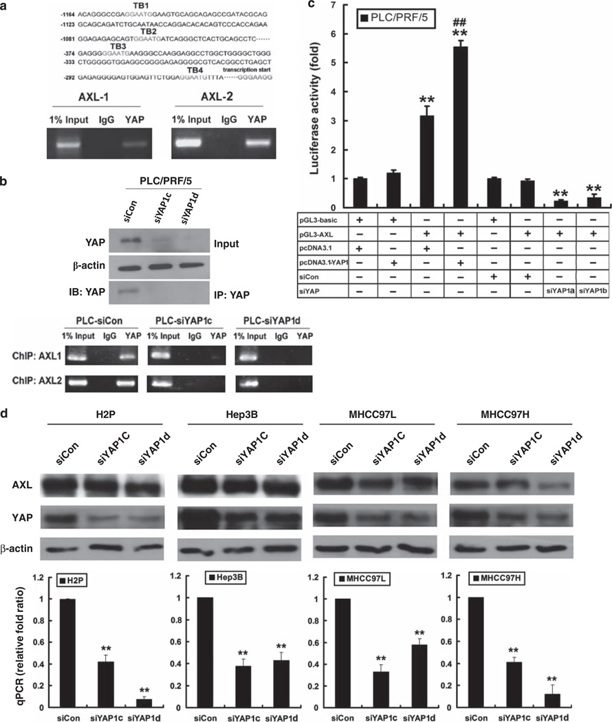
Transcriptional activation of AXL receptor kinase by YAP. (a) ChIP assay in MIHA-YAP1 cells. Four putative TB (TB1–4) were found in the AXL promoter region (upper panel). AXL promoter-specific PCR covering the TEAD-binding sequences TB1–TB2 or TB3–TB4 was conducted (lower panel). A PCR band was detected using the anti-YAP antibody, but not the IgG control, suggesting that YAP activates AXL through TEAD transcription factor. (b) Immunoblotting and ChIP assay in PLC-siYAP1cells. Immunoprecipitation (IP) of YAP from PLC-siYAP1c and PLC-siYAP1d cells, followed by immunoblotting (IB) with anti-YAP antibody, showed decreased levels of YAP after siRNA treatments, when compared with the siCon control. Total lysate input and β-actin were included as references. ChIP assay of the same immunocomplexes showed decreased signal of AXL promoter sequences in the PLC-siYAP1cells. (c) Luciferase reporter assay in PLC/PRF/5 cells with YAP overexpression or underexpression. pGL3-basic vector-transfected cells were included as controls. PLC/PRF/5 cells transfected with pGL3-AXL showed significantly elevated reporter activity compared with controls; meanwhile, cells co-transfected with pcDNA3.1-YAP1 exhibited further enhanced luciferase activity. For PLC/PRF/5 cells with YAP underexpression, siCon-transfected cells were included as controls. PLC/PRF/5 cells transfected with pGL3-AXL and siYAP1 showed significantly decreased reporter activity compared with controls. Cells were harvested 24 h after transfection for measurement of luciferase activity in all experiments. Total amounts of DNA and RNA were kept constant. All experiments were conducted in triplicate and repeated three times. Data are shown as mean ± 2 × s.e.m. (** compared with controls, P < 0.001; ## compared with pcDNA3.1 and pGL3-AXL co-transfectant, P < 0.01). (d) Reduced expression of AXL after transient knockdown of YAP1 in four different HCC cell lines—H2P, Hep3B, MHCC97L and MHCC97H. YAP1 Stealth RNAi siRNAs (#HSS115942 for siYAP1c and #HSS115944 for siYAP1d) were used to knock down YAP1 expression in HCC cells. Immunoblotting of YAP and AXL proteins (upper panel) and quantitative PCR assay of AXL transcript (lower) were carried out according to the procedures described above **P < 0.01.

