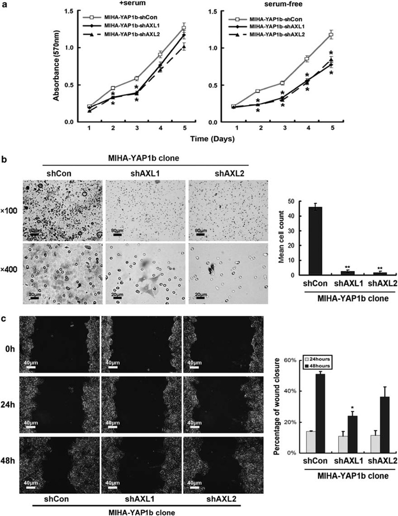
Figure 6.
AXL is required for cell viability and invasion in theMIHA-YAP1 clone. (a)MTT analysis showed that downregulation of AXL by AXL short hairpin RNA transient transfection (MIHA-YAP1b-shAXL1 and MIHA-YAP1b-shAXL2) decreased cell viability and growth in MIHA-YAP1b cells in the presence of serum compared with vector-transfected controls (MIHA-YAP1b-shCon) (left panel, comparison between transfectant and vector control, each day, using Student’s t-test; *P < 0.05 on days 2 and 3). In serum-free medium, MIHA-YAP1b-shAXL1 and MIHA-YAP1b-shAXL2 showed impaired cell viability and growth compared with MIHA-YAP1b-shCon (right panel, comparison as described above; *P < 0.05 on days 2, 3, 4 and 5). All experiments were conducted in triplicate in three independent experiments. Data are shown as mean ± 2 × s.e.m. (b) Matrigel invasion assay showed thatMIHA-YAP1b-shAXL1 or MIHA-YAP1b-shAXL2 could not penetrate the matrigel membrane within 72 h of culture. Representative fields of invasive cells on the membrane are shown (left panel). Penetrated cells were stained with 0.1% crystal violet. Upper panel: × 100, scale bar: 80 µm; lower panel: × 400, scale bar: 20 µm. MIHA- YAP1b-shAXL cells showed significantly decreased number of penetrated cells (right panel) **P < 0.01. (c) AXL affected cell migration in YAP-overexpressing MIHA cells. MIHA-YAP1b-shAXL1 and MIHA-YAP1b-shAXL2 cells showed moderately slowed migration during 48 h compared with MIHA-YAP1b-shCon cells (left panel). Magnification: × 200, scale bar: 40 µm. Compared with control cells, percentages of wound closure were moderately decreased in MIHA-YAP1b-shAXL cells (right panel) *P < 0.05.

