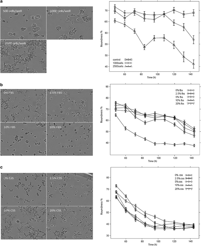
Figure 1.
Factors supporting the invasive switch of acini formed by PC-3 cells in organotypic 3-D culture. (a) Increasing cell density promotes conversion of differentiated acini into invasive structures (phase-contrast images from live cell imaging; left panel). Disintegration of spheroids and invasive properties measured as loss of roundness by automated image analysis software ACCA (right panel; measured from days 4–7 of culture). (b) Addition of >2.5% FBS supports differentiation and suppresses invasive transformation of acini in 3-D. (c) FBS deprived of lipids by charcoal-stripping (CSS) fails to support differentiation even at 20%.

