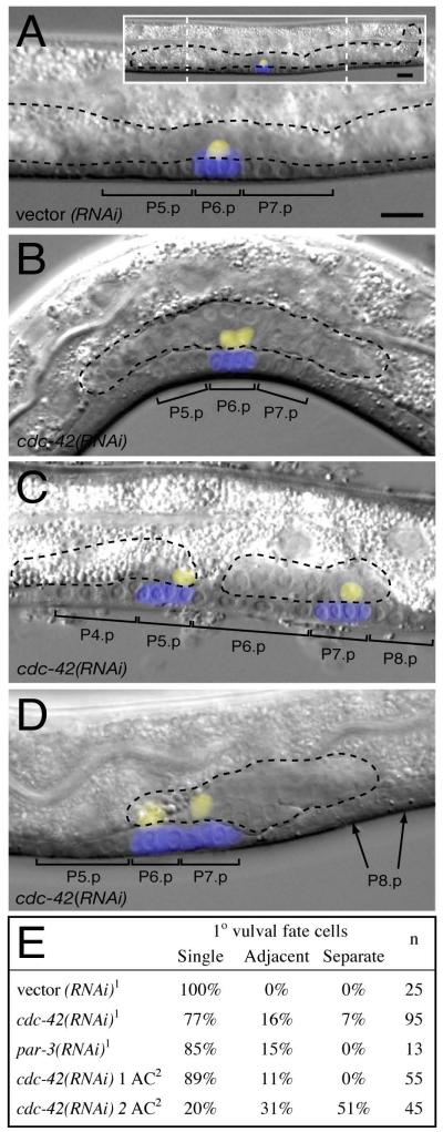
Figure 2. cdc-42(RNAi) results in multiple ACs, adjacent 1° VPC fates and gonad splitting.
Overlays of Nomarski images of L3 Pn.pxx stage animals with projections of zmp-1::YFP expression (yellow) marking the Anchor Cell (AC) and egl-17::CFP expression (blue) marking 1° fate VPCs. Brackets indicate the descendants of induced VPCs and black dotted outlines the morphology of the gonad.
(A) wild-type with extended gonad (inset), a single AC, and three induced VPCs, one of which (P6.p) adopts the 1° fate (marked by egl-17::CFP).
(B-D) cdc-42(RNAi) animals with (B) two touching ACs and a single 1° fate VPC, (C) two ACs in separate gonad fragments and two, non-adjacent, 1° fate VPCs, (D) two separated ACs in an intact, but short, gonad and two adjacent 1° fate VPCs. . VPCs adjacent to 1° fate VPCs sometimes fail to adopt vulval fates (arrows in D), perhaps because of a gap between the VPCs.
Anterior, left; ventral, down; scale bar, 10μm
(E) Table of 1° vulval fate phenotypes
11° fate scored using arIs92[egl-17::CFP::lacZ] (Yoo et al., 2004)
21° fate scored using syIs59[egl-17:CFP] (Inoue et al., 2002)
AC: Anchor Cell

