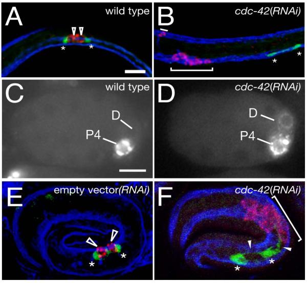
Figure 5. Germ cells adopt muscle fates and are not associated with SGPs in cdc-42(RNAi) animals.

SGPs (ehn-3A::GFP, green), P-granules (anti-PGL-1 (Kawasaki et al., 1998), red in (A,B,E,F), white in (C,D)), and body wall muscles (NE8/4C6.3 marker (Goh and Bogaert, 1991), blue). (A) wild-type L1 gonad primordium with polar SGPs (asterisks) and central PGCs (unfilled arrowheads) (B) cdc-42(RNAi) L1 animal with P-granules (red, brackets) in muscle cells (blue), and SGPs (green) lacking associated PGCs (no P-granule containing cells).
(C) wild-type embryo after the division of P3, the P granule marker pgl-1::GFP (Cheeks et al., 2004) is only in the germline precursor P4. (D) cdc-42(RNAi) embryo with P-granules in both P4 and its sibling D.
(E) wild-type 3-fold embryo; SGPs are associated with PGCs (F) cdc-42(RNAi) embryo; SGPs extend processes that appear to be directed towards the PGL-1 staining cells (filled arrowheads).
Anterior, left; scale bar ,10μm.
