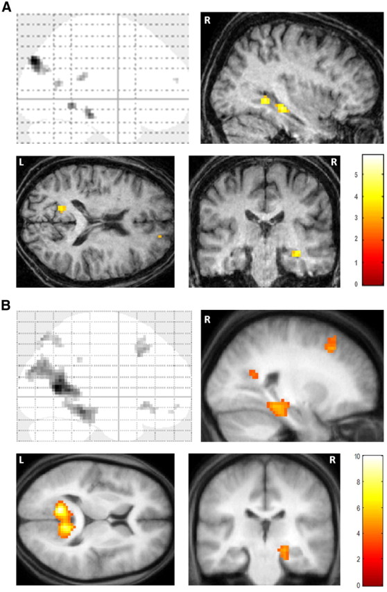
Figure 4.

fMRI results. A, Brain areas more active for constructing fictitious scenes compared with imagining single acontextual objects in patient P01. Upper left, Sagittal image from a “glass brain,” which enables one to appreciate activations at all locations and levels in the brain simultaneously. Activations are shown on sagittal (upper right), axial (lower left), and coronal (lower right) images from P01's structural scan at a threshold of p < 0.001 (whole brain, uncorrected). The color bar indicates the z-scores associated with each voxel. L, Left side of the brain, R, right side of the brain. B, The same contrast in 21 healthy participants [data from Hassabis et al. (2007b)] shown on the averaged structural MRI scan of those participants.
