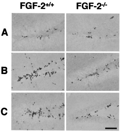
Figure 1.
BrdUrd-positive cells in the medial dentate gyrus of FGF-2+/+ and FGF-2−/− mice after brain injury. After kainic acid injection, MCAO or no injury (control), BrdUrd was injected 6, 7, and 8 days later (to label dividing cells), and animals were killed on day 9. Few BrdUrd-labeled cells were detected in the untreated FGF-2+/+ and FGF-2−/− mice (A). After kainic acid injection (B) or MCAO (C), greater cell proliferation was detected in a region corresponding to the subgranular layer in FGF-2+/+ littermates. Immunohistochemistry was performed on free-floating 50-μm coronal sections pretreated by denaturing DNA (see Materials and Methods). (Scale bar, 100 μm.)

