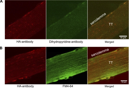
Fig. 5.
Immunofluorescence detection of surface-exposed HA tag and colocalization with T-tubules. A: isolated nonpermeabilized FDB fibers from HA-GLUT4-GFP Tg mice were stained with anti-HA (red) and dihyropyridine receptor (green) antibodies. Cells were stimulated with insulin for 30 min and stained with mouse anti-HA and rabbit anti-dihyropyridine antibodies for 15 min at RT. After fixation, primary antibodies were detected with goat anti-mouse-Alexa 594 and goat anti-rabbit-Alexa 488 secondary antibodies. Note high level of colocalization at T-tubules. Bar, 10 μm. Images shown are representative cells from 2 experiments. B: colocalization of HA antibody (red) and lipid marker FM4-64 (pdseudocolored green) at T-tubules of isolated nonpermeabilized FDB fibers. Cells were stimulated with insulin for 30 min and stained with anti-HA antibody for 15 min, then fixed and labeled with Alexa 594-conjugated secondary antibody. Images shown are representative cells from 3 experiments.

