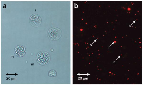
Figure 2.

Images of chromosome preparation and staining. (a) Swollen cells in metaphase (m) and interphase (i) stained with Turck’s solution (Step 7). (b) Released single chromosomes (s) and interphase nuclei (i) after treating the swollen cells with polyamine isolation buffer containing Triton X-100, and staining with propidium iodide (Step 11).
