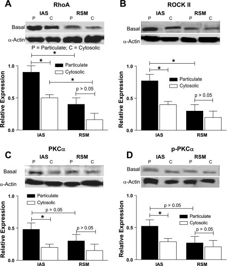
Fig. 3.
Western blot data showing significantly higher levels of expressions of RhoA/ROCK II, PKC-α, and phosphorylated PKC-α (p-PKC-α) (A–D) in the smooth muscle tissues of the IAS vs. RSM, especially in the particulate fractions, in the basal state (*P < 0.05). In addition, RhoA levels are higher in the cytosolic fractions of the IAS vs. the RSM (*P < 0.05). The significance of the latter observations remains to be determined. The levels of PKC-α (C) and p-PKC-α (D), although higher, are significantly less compared with RhoA/ROCK II. Data show no significant differences between the particulate and cytosolic fractions of the RSM (P > 0.05).

