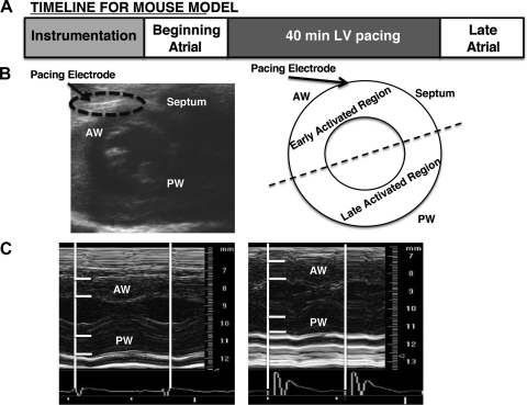
Fig. 1.
Pacing preparation and experimental protocol. A: experimental design for mouse model of ventricular dysynchrony. Briefly, a unipolar pacing electrode was inserted into the left ventricle (LV) of the mouse. Successful implantation of the electrode was visualized by the 2-dimensional echo images (B) and the induction of ventricular dysynchrony was determined by M-mode echocardiography analysis of anterior and posterior wall changes in wall thickness during the cardiac cycle (C). Tissue samples were collected from the early-activated region and late-activated regions from all hearts and used for biochemical analysis. AW, anterior wall; PW, posterior wall.

