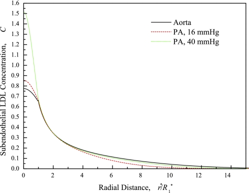
Fig. 10.
Distribution of LDL concentration (C) in the subendothelial intima, normalized by the plasma LDL concentration, as a function of r at 10 min LDL circulation time for the normotensive aorta (solid line) and the PA with normal (16 mmHg, dashed line) and elevated (40 mmHg, dotted line) transmural pressure. Parameters: L*1 = 1 μm (23); media LDL distribution volumes γ2 estimated from HRP values and molecular radii: 0.025 (aorta), 0.074 (PA); PA parameters at 16 and 40 mmHg same except for L*pt(L*pnj) = 1.60 × 10⁻⁷ (3.35 × 10⁻⁷) cm·s−1·mmHg−1 at ΔP* = 40 mmHg (45). L*pnj is 41% lower than at 16 mmHg (24, 45).

