Fig. 2.
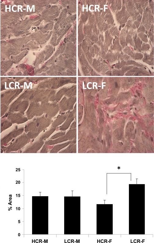
Top: representative light micrographs of Verhoeff van Gieson-stained sections of the LV showing pink-stained collagen fibers in the myocardial interstitium. Bottom: bar graph showing the results of a quantitative morphometric analysis indicating increased interstitial fibrosis in LCR-F compared with HCR-F (n = 5 rats/group). *P < 0.05.

